
Research in to Brain Tumours
Project overview
The Glioma Team at the ICR is dedicated to turning biological discoveries into improved clinical outcomes for children with brain tumours. In order to continue building on their progress, the team needs ongoing support in the characterisation of childhood glioma, and the testing of new drugs in laboratory models. Support from the Ollie Young Foundation is instrumental in helping them to achieve this.
The research Professor Chris Jones’s team is undertaking generates a wealth of complex information. This information can be very illuminating – it can shed light on the characteristics of childhood glioma, and, in turn, promising new drugs can be identified. The key to making the most of this treasure-trove of data is to ensure that it can be efficiently and effectively analysed. Yura Grabovska’s expertise helps the team to do this by ensuring that these datasets can be visualised and interpreted. He also has a passion for empowering his colleagues to better understand their data themselves so they can ask the most meaningful questions of it.
A key area of research
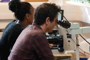
The team has ongoing collaborations with local, national, and international hospitals whereby they receive surgical and biopsy material from children with glioblastoma and DIPG. They receive samples of tumour, in excess of that required for routine diagnosis. Upon receiving a sample, the team gently breaks the tissue into single cells, and grows them in a dish under a controlled environment (‘in vitro’). In addition, they implant cells into the brain of mice, in order to form tumours which resemble the human disease (‘in vivo’). Whilst these models are being established, they also carry out in-depth molecular analysis in order to understand the specific genetic alterations driving that child’s individual tumour.
Once they have enough cells, large panels of drugs are tested in vitro, with the aim of identifying drugs which kill the cancer cells from a given subtype of the disease. Promising drugs identified in this way are later tested again in vivo in order to provide data which may lead to future clinical trials. Putting these complicated data together is one of the most challenging aspects of what the team does. Yura has specific expertise in handling these large datasets, integrating them, and presenting the data in such a way as the biologists can interpret them. This helps them to gain insight into which drugs should be prioritised for further work based upon the individual molecular ‘signature’ of tumours. In addition to this, he also provides a range of analytical support across our whole research programme. He is invaluable to the team.
How the Ollie Young Foundation is supporting this vital research
Support from the foundation is funding Yura’s vital research within the team. In addition to funding his salary, the foundation is also contributing a budget for both molecular screening and drug screening costs. This rounded support is enabling experiments to be undertaken to generate the detailed datasets that Yura dedicates himself to interpreting.
Research Insight

This image shows a heatmap of patient-derived cell lines being screened with a large panel of potential cancer therapeutic agents. The columns are the drugs, and the rows are different patient-derived cell line models. On the left, the green/grey/blue refers to different histone gene mutations in high-grade glioma and DIPG. Drug screening is done by testing different, escalating concentrations of each drug. The ‘hotter’ or more yellow the result is, the lower the concentration of drug is needed to stop the cancer cell being able to survive in culture, meaning it is more likely to be a potentially effective drug in that cancer. These results were generated by the team and are currently in the process of being published. An open-access version of the paper is available here.

This image is a visual representation of almost 2000 tumour profiles derived from our own team’s work as well as other published data. The tumour sub-groups shown include low-grade and high-grade gliomas of different types, affecting children and adults. When we receive a new patient tumour sample, we can use a method called ‘DNA methylation microarray’ to generate a genome-wide profile of the tumour and we can then project that sample onto this map which gives a useful representation of the tumour type. This visualisation is a standard approach in the field and means that we can very easily and meaningfully share information with the group and collaborators about different tumours we are working on together. As well as this, the tumours landing in specific groups suggests to us various aspects of their biology which we can explore further. An example of this plot is in the paper linked above.